Multiple timescale dynamics of conductance-based models of brainstem locomotor neurons
A. K. Thomas & J. E. Rubin (2026). arXiv:2603.11467, under review at SIAM Life Sciences.
The pedunculopontine nucleus (PPN) is a heterogeneous brainstem locomotor hub implicated in Parkinson’s disease and a target for deep brain stimulation (DBS). Existing PPN models are either population-level firing-rate models or small conductance sets calibrated to a single cell type, and they fail to reproduce the distinctive single-cell dynamics observed experimentally.
We developed three single-compartment Hodgkin–Huxley-type models from the ground up for the cholinergic (C), cholinergic with low-threshold Ca2+ spikes (CT), and non-cholinergic (NC) PPN cell classes. Each model reproduces its class’s stimulus-dependent responses, including post-inhibitory rebound, transient low-threshold spiking, and gamma-band (30–90 Hz) oscillations. The models combine Na+, K+, P/Q- and T-type Ca2+, A-type K+, and Ca2+-activated K+ currents with intracellular calcium dynamics.
Approach
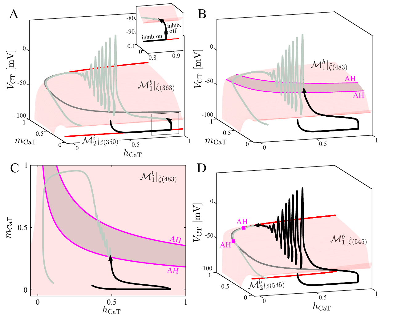
We nondimensionalized each model to separate the variables into fast (membrane potential, fast gating), slow, and, in the CT model, superslow classes (slow gating and calcium). We then used geometric singular perturbation theory and bifurcation analysis of the fast subsystem, parameterized by the slower variables, to explain each transient response. Unlike most analyses of this type, capturing some of these responses required accounting for the voltage dependence of the gating timescales themselves.
Key results
- C model: the delay before firing resumes after inhibition arises from slow A-current (IA) gating.
- CT model: T-type Ca2+ kinetics are the dominant control of post-inhibitory rebound oscillations, which are organized by three-timescale dynamics.
- NC model: P/Q- and T-type Ca2+ conductances together shape high-voltage gamma oscillations and low-threshold spikes; spiking initiates at a fast-subsystem SNIC bifurcation.
- Prediction: under an untested post-inhibitory facilitation protocol, only the cell classes expressing T-type Ca2+ current (CT and NC) show facilitation, each with a distinct signature.
Why it matters
The models tie distinct firing phenotypes to specific ion channels. This is the level at which pharmacological and stimulation-based interventions act, and it suggests how inhibitory input from the substantia nigra pars reticulata and DBS-like stimulation may differentially shape output across PPN cell types in motor circuits.